Aller au contenu principal

Thèse d'Habib Murtaza

Le 30 mars 2022

Towards improved laboratory X-ray tomography imaging: optimization of acquisition parameters and use of photon-counting detectors.

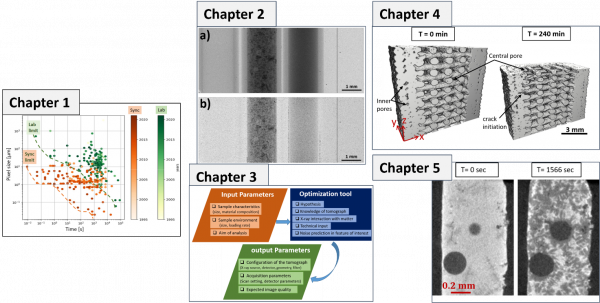
This study deals with developments to increase the possibilities offered by laboratory X-ray computed tomography in material science by focusing on contrast enhancement and on time resolution aspect. First, the feasibility of using a new generation photon-counting detector (PCD) in lab-CT was evaluated. The characterization of the standard imaging performances and the spectral capabilities of four PCDs were carried out and compared to a standard flat-panel detector.  The potential of PCD towards spectral and single-shot K-edge imaging was investigated. Second, a model-based optimization strategy is developed to define the suitable CT scanning parameters for dynamic in situ acquisitions with an image quality allowing qualitative or quantitative analysis. The model is based on three modules: modelling noise in the feature of interest, X-ray absorption simulation tool, and the screening algorithm that outputs the different possible scanning configurations associated with the probability of detection of the interested feature size for each configuration.  A real-time in-situ test with sub-minute temporal resolution was performed with the experimentally optimized CT set-up as an application aspect of the thesis. The experimental configuration is confronted with the proposed optimization model configurations, which were found to be in-line with the chosen setup. The application corresponds to the real-time monitoring of microstructural evolution of 3D printed cellulose parts during air-drying phenomena with qualitative and quantitative analysis.  It illustrates the quantitative characterization capabilities of lab-CT for high-speed in-situ imaging.
 
Keywords: X-ray tomography, In-situ tomography, Time-resolved imaging, Photon-counting detector, Parameter optimization, Image analysis

Jury

Daniel Bellet, Professor, Grenoble INP, (Examiner)
Eric Maire, Research Director, CNRS, INSA Lyon, (Reviewer)
Dominique Bernard, Research Director, CNRS, ICMCB Bordeaux, (Reviewer)
Pierre Dumont, Professor, INSA Lyon, (Examiner)
Barbara Fayard, CEO Novitom, Grenoble, (Member)
Sabine Rolland Du Roscoat, Maître de conférences, Université Grenoble Alpes, 3SR, (Thesis Director)
Pierre Lhuissier, Chargé de Recherche, CNRS, Grenoble INP, SIMAP, (Thesis Co-director)
Luc Salvo, Professor, Grenoble INP, SIMAP, (Thesis Co-director)
Partenaires
partenaires.jpg

Date

Le 30 mars 2022
Complément date
9h30

Localisation

Complément lieu
Amphi Kilian, 1381 Rue de la Piscine, 38610 Gières
Zoom link for the audience

habib.jpg

Figure caption : Thesis outline : Pixel size versus scan time illustrating the state of the art for in-situ X-ray synchrotron and laboratory computed tomography (Chapter 1). Result from Photon-counting detector: Standard flat-field corrected radiograph of the (I-BaSO4-H2O) phantom recorded with Eth_low = 5.0 keV. (b) KES flat-field corrected radiograph of the (I-BaSO4-H2O) phantom recorded with using Eth_low = 28 keV and Eth_high =38 keV (Chapter 2). Overview of the optimization model to estimate the optimal CT scanning acquisition parameters for dynamic in-situ scans (Chapter 3).  3D rendered cross-sectional view of reconstructed X-ray tomographic scans at different drying time. Application for air-drying monitoring of 3D printed part was performed by fast laboratory X-Ray microtomography (Chapter 4). 2D reconstructed slices of Al-Si-Cu alloy during the solidification experiment performed by fast laboratory X-Ray microtomography (Chapter 5).

Publié le 29 mars 2022

Mis à jour le 29 mars 2022