- Share
- Share on Facebook
- Share on LinkedIn
On March 30, 2022
Towards improved laboratory X-ray tomography imaging: optimization of acquisition parameters and use of photon-counting detectors.
Keywords: X-ray tomography, In-situ tomography, Time-resolved imaging, Photon-counting detector, Parameter optimization, Image analysis
Jury
Dominique Bernard, Research Director, CNRS, ICMCB Bordeaux, (Reviewer)
Pierre Dumont, Professor, INSA Lyon, (Examiner)
Barbara Fayard, CEO Novitom, Grenoble, (Member)
Sabine Rolland Du Roscoat, Maître de conférences, Université Grenoble Alpes, 3SR, (Thesis Director)
Pierre Lhuissier, Chargé de Recherche, CNRS, Grenoble INP, SIMAP, (Thesis Co-director)
Luc Salvo, Professor, Grenoble INP, SIMAP, (Thesis Co-director)

Date
9h30
Localisation
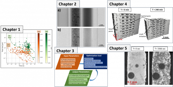
Figure caption : Thesis outline : Pixel size versus scan time illustrating the state of the art for in-situ X-ray synchrotron and laboratory computed tomography (Chapter 1). Result from Photon-counting detector: Standard flat-field corrected radiograph of the (I-BaSO4-H2O) phantom recorded with Eth_low = 5.0 keV. (b) KES flat-field corrected radiograph of the (I-BaSO4-H2O) phantom recorded with using Eth_low = 28 keV and Eth_high =38 keV (Chapter 2). Overview of the optimization model to estimate the optimal CT scanning acquisition parameters for dynamic in-situ scans (Chapter 3). 3D rendered cross-sectional view of reconstructed X-ray tomographic scans at different drying time. Application for air-drying monitoring of 3D printed part was performed by fast laboratory X-Ray microtomography (Chapter 4). 2D reconstructed slices of Al-Si-Cu alloy during the solidification experiment performed by fast laboratory X-Ray microtomography (Chapter 5).
- Share
- Share on Facebook
- Share on LinkedIn